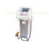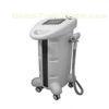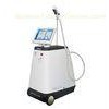


Mini 3 in 1 3 M Ultrasonic Skin Microdermabrasion Equipments home use Machine
1. Functions:
1. Electroporation
2. 3 M Ultrasonic
3. Diamond Dermabrasion
2. Electroporation Working Theory
Electroporation is a mechanical method used to introduce polar molecules into a host cell through the cell membrane. In this procedure, a large electric pulse temporarily disturbs the phospholipid bilayer, allowing molecules like DNA to pass into the cell.
3 in1 Electroporation
3. Technical parameters.
Power Input: 110V/60Hz or 220V/50Hz
Max. Power: 30 W
Temperature range: 10-42
4. Accessories
Main unit 1 pc
Electroporation Treatment probe 2 pcs
3M Ultrasonic Treatment Probe 2 pcs
Anode plate 1 pc
Treatment cable 1 pc
Power Cord 1 Pc
Manual 1 pc
Fuse 2 pc
Paper Carton 1 pc
Packing Foam 1 set
5. Background
Many research techniques in molecular biology require a foreign gene or protein material to be inserted into a host cell. Since the phospholipid bilayer of the plasma membrane has a hydrophobic exterior and a hydrophobic interior (Fig. 1), any polar molecules, including DNA and protein, are unable to freely pass through the membrane.
Figure 1. Diagram of the Phospholipid Bilayer. This image shows the chemical components of the plasma membrane. The polar head groups face outward while the hydrophobic tail groups face inward and interact with one another to hold the membrane together. Polar molecules cannot pass through this membrane without external aid.
Many methods have been developed to surpass this barrier and allow the insertion of DNA and other molecules into the cells to be studied. One such method is electroporation.
The concept of electroporation capitalizes on the relatively weak nature of the phospholipid bilayer's hydrophobic/hydrophilic interactions and its ability to spontaneously reassemble after disturbance. Thus, a quick voltage shock may disrupt areas of the membrane temporarily, allowing polar molecules to pass, but then the membrane may reseal quickly and leave the cell intact.
Procedure
The host cells and the molecules to be inserted into these cells are suspended in solution. The electroporation apparatus is typically commercially produced and purchased, but the basic process inside such an apparatus may be represented in a schematic diagram
Figure 2. Diagram of the basic circuit setup of the electroporation apparatus. This diagram shows the basic electric circuit that provides the voltage for electroporation.
When the first switch is closed, the capacitor charges up and stores a high voltage. When the second switch is closed, this voltage discharges through the liquid of the cell suspension. Typically, 10, 000-100, 000 V/cm (varying with cell size) in a pulse lasting a few microseconds to a millisecond is necessary for electroporation. This electric pulse disturbs the phospholipid bilayer of the membrane and causes the formation of temporary aqueous pores. The electric potential across the membrane of the cell simultaneously rises by about 0.5-1.0 V so that charged molecules (such as DNA) are driven across the membrane through the pores in a manner similar to electrophoresis (Fig 3)
Figure 3. Graphic representation of plasmids containing a foreign DNA insert passing through temporary aqueous pores in the plasma membrane. The actual entry of
Mini 3 in 1 3 M Ultrasonic Skin Microdermabrasion Equipments home use Machine